
Peroneal tendonitis is an inflammation of the peroneal tendons‚ often caused by overuse or injury‚ requiring targeted exercises for effective rehabilitation and pain relief.
1.1 Overview of Peroneal Tendonitis
Peroneal tendonitis involves inflammation of the peroneal tendons‚ which run along the outer side of the ankle. These tendons play a crucial role in stabilizing the ankle and facilitating movements like eversion and dorsiflexion. The condition often arises from repetitive stress‚ overuse‚ or acute injuries‚ leading to pain and swelling on the outside of the ankle. If left untreated‚ it can progress to chronic tendinopathy‚ causing prolonged discomfort and limited mobility. Early intervention‚ including rest‚ icing‚ and targeted exercises‚ is essential to promote healing and restore function. Exercises such as ankle eversion‚ heel raises‚ and single-leg balance are commonly recommended to strengthen the surrounding muscles and improve tendon resilience. Proper rehabilitation can prevent further complications and ensure a successful recovery.
1.2 Importance of Exercises in Rehabilitation
Exercises play a vital role in the rehabilitation of peroneal tendonitis‚ promoting healing and restoring function. They help improve blood flow‚ reduce inflammation‚ and strengthen the muscles surrounding the tendons‚ preventing imbalances. Strengthening and stretching exercises enhance tendon resilience‚ reducing the risk of re-injury. Additionally‚ exercises improve ankle mobility and proprioception‚ which are essential for overall stability. A structured exercise program can address pain‚ swelling‚ and limited mobility‚ ensuring a faster and more effective recovery. By incorporating dynamic and static stretches‚ as well as resistance training‚ patients can regain strength and confidence in their ankle. Consistency in performing these exercises is key to achieving long-term benefits and preventing future complications.
Pathophysiology of Peroneal Tendonitis
Peroneal tendonitis arises from inflammation and degeneration of the peroneal tendons‚ often due to repetitive stress or overuse‚ leading to pain and impaired tendon function.
2.1 Anatomy of the Peroneal Tendons
The peroneal tendons are two fibrous cords‚ the peroneus longus and peroneus brevis‚ located on the lateral side of the lower leg and foot. They originate from the fibula and attach to the foot bones‚ stabilizing the ankle and facilitating eversion‚ the outward movement of the foot. These tendons run in a groove behind the lateral malleolus‚ protected by a fibrous sheath. Their primary function is to support the lateral ankle and enable movements essential for walking and balance. Understanding their anatomy is crucial for diagnosing injuries and developing targeted exercises for rehabilitation. Proper alignment and mobility of these tendons are vital for preventing tendonitis and ensuring optimal lower limb function.
2.2 Causes and Risk Factors
Peroneal tendonitis typically arises from overuse‚ repetitive stress‚ or sudden increases in physical activity‚ leading to inflammation and degeneration of the tendons. Risk factors include weak peroneal muscles‚ tight calf muscles‚ and participation in high-impact sports or activities that involve repetitive ankle movements. Improper footwear‚ lack of arch support‚ and training on uneven surfaces can also contribute. Anatomical issues‚ such as a low arch or fibular malalignment‚ may predispose individuals to this condition. Obesity and previous ankle injuries or sprains further increase the likelihood of developing peroneal tendonitis. Addressing these factors through targeted exercises‚ such as heel raises and calf stretches‚ can help mitigate symptoms and prevent further damage;
Symptoms of Peroneal Tendonitis
Pain on the outside of the ankle‚ swelling‚ and limited mobility are common symptoms. Activities may worsen discomfort‚ with tenderness and warmth around the affected area.
3.1 Common Pain Locations
Peroneal tendonitis typically causes pain on the outside of the ankle‚ particularly behind the lateral malleolus (the bony prominence on the outer ankle). Pain may also radiate along the tendon’s path‚ extending upward along the lower leg or downward toward the foot. The discomfort is often most pronounced during activities that involve ankle movement‚ such as running‚ walking on uneven surfaces‚ or climbing stairs. Tenderness and swelling may be present in the affected area‚ and patients might experience warmth or redness around the tendon. In severe cases‚ pain can persist even at rest‚ making it difficult to bear weight or perform daily activities. Early recognition of these symptoms is crucial for effective management and rehabilitation.
3.2 Swelling and Limited Mobility
Swelling is a common symptom of peroneal tendonitis‚ often accompanied by limited mobility in the affected ankle. The inflammation causes fluid retention‚ leading to visible swelling around the lateral malleolus (outer ankle bone). This swelling can press on nearby tissues‚ increasing pain and stiffness. Patients may experience difficulty moving the ankle‚ particularly during inversion (turning inward) and eversion (turning outward) movements. The restricted range of motion can hinder daily activities like walking or climbing stairs. In severe cases‚ the swelling may extend along the tendon’s path‚ further reducing mobility. Prolonged limited movement can lead to muscle weakness and reduced flexibility‚ emphasizing the need for early intervention to restore function and prevent chronic issues. Addressing swelling and improving mobility are key focuses in the rehabilitation process.

Diagnostic Criteria
Diagnosis involves physical examination‚ patient history‚ and imaging to confirm peroneal tendon inflammation‚ ensuring accurate identification of symptoms and appropriate treatment plans for rehabilitation.
4.1 Physical Examination
A physical examination is crucial for diagnosing peroneal tendonitis. It involves palpation to identify tenderness along the peroneal tendons‚ located behind the lateral malleolus. Swelling and warmth in the affected area are common findings. The physician may perform specific tests‚ such as the provocation test‚ where resistance is applied during ankle eversion to replicate pain. Range of motion assessment and gait evaluation can reveal limitations or abnormalities. The examiner may also check for instability or subluxation of the tendons. Patient history‚ including recent activities or injuries‚ is essential to guide the examination. These findings help differentiate peroneal tendonitis from other ankle conditions‚ ensuring an accurate diagnosis and appropriate treatment plan.
4.2 Imaging Studies
Imaging studies are essential for confirming the diagnosis of peroneal tendonitis and ruling out other conditions. Magnetic Resonance Imaging (MRI) is the most commonly used modality‚ providing detailed images of the tendons‚ muscles‚ and surrounding soft tissues. It can reveal tendon thickening‚ inflammation‚ or tears. Ultrasound is another effective tool‚ offering real-time visualization of the tendons and guiding injections if needed. X-rays are typically used to exclude fractures or bony abnormalities. In some cases‚ a musculoskeletal ultrasound may be employed to assess tendon mobility and detect subluxation or dislocation. These imaging techniques help identify the extent of tendon damage and guide appropriate treatment options‚ ensuring a comprehensive approach to managing peroneal tendonitis.
Treatment Options for Peroneal Tendonitis
Treatment for peroneal tendonitis often combines rest‚ physical therapy‚ and anti-inflammatory medications. Surgery may be needed for severe cases or tendon tears‚ ensuring proper recovery and functionality.
5.1 Conservative Management
Conservative management for peroneal tendonitis focuses on reducing inflammation and promoting tendon healing. Initial treatment often involves the RICE method: rest‚ ice‚ compression‚ and elevation. Nonsteroidal anti-inflammatory drugs (NSAIDs) may be prescribed to alleviate pain and swelling. Physical therapy plays a central role‚ incorporating exercises such as calf stretches‚ single-leg balance drills‚ and wobble board exercises to improve strength and stability. Gentle mobilization of the ankle and calf muscles is also recommended to restore range of motion. Orthotics or supportive footwear can reduce stress on the tendons. Patients are encouraged to avoid high-impact activities during the acute phase and gradually reintroduce them as symptoms improve. This approach aims to avoid surgical intervention by addressing the root causes of tendon irritation and strengthening the surrounding tissues.
5.2 Surgical Interventions
Surgical interventions for peroneal tendonitis are typically considered when conservative treatments fail to alleviate symptoms. Common procedures include tendon debridement‚ where damaged or inflamed tissue is removed‚ or tendon transfer to redistribute stress. In severe cases‚ such as complete tendon tears‚ surgical repair may be necessary. These procedures aim to restore tendon function and eliminate pain. Recovery often involves a period of immobilization‚ followed by gradual rehabilitation to regain strength and mobility. Surgery is generally a last resort‚ reserved for patients who do not respond to non-invasive treatments. The decision to operate is made in consultation with orthopedic specialists‚ ensuring the best possible outcome for long-term tendon health.

Exercise Program for Peroneal Tendonitis
A comprehensive exercise program for peroneal tendonitis focuses on stretching‚ strengthening‚ and improving balance to promote healing‚ reduce pain‚ and restore ankle function effectively.
6.1 Stretching Exercises
Stretching exercises are essential for managing peroneal tendonitis‚ as they improve flexibility and reduce tightness in the tendons and surrounding muscles. One effective stretch is the calf stretch‚ which targets the muscles connected to the peroneal tendons. To perform this‚ stand facing a wall with one hand on the wall for balance. Step one foot back about a foot‚ keeping your heel on the ground‚ and gently bend the front knee. Hold for 15-30 seconds and repeat on the other side. Another beneficial stretch is the towel stretch‚ where you sit on the floor with your injured leg extended. Loop a towel around your foot and gently pull it toward you to feel a stretch in the calf and peroneal tendons. Hold for 15-30 seconds and repeat 3 times. Regular stretching can help alleviate symptoms and promote healing.
6.2 Strengthening Exercises
Strengthening exercises are crucial for rehabilitating peroneal tendonitis‚ as they enhance muscle stability and reduce tendon stress. Heel raises are an excellent option‚ targeting the peroneal tendons and calf muscles. Stand with feet shoulder-width apart‚ hold onto a sturdy object for balance‚ and slowly raise your heels off the ground before lowering back down. Perform 3 sets of 12-15 repetitions. Single-leg balance exercises also strengthen the muscles surrounding the peroneal tendons. Stand on the affected leg‚ keeping the other foot lifted‚ and hold for 20-30 seconds‚ switching legs afterward. Resistance band exercises‚ such as eversion (rotating the foot outward)‚ can be done by looping a band around the foot and pulling it sideways. Heel raises on a step further target the tendons and calf muscles. These exercises should be done without pain‚ focusing on muscle fatigue rather than discomfort‚ and repeated 2-3 times daily for optimal results.
6.3 Balance and Proprioception Training
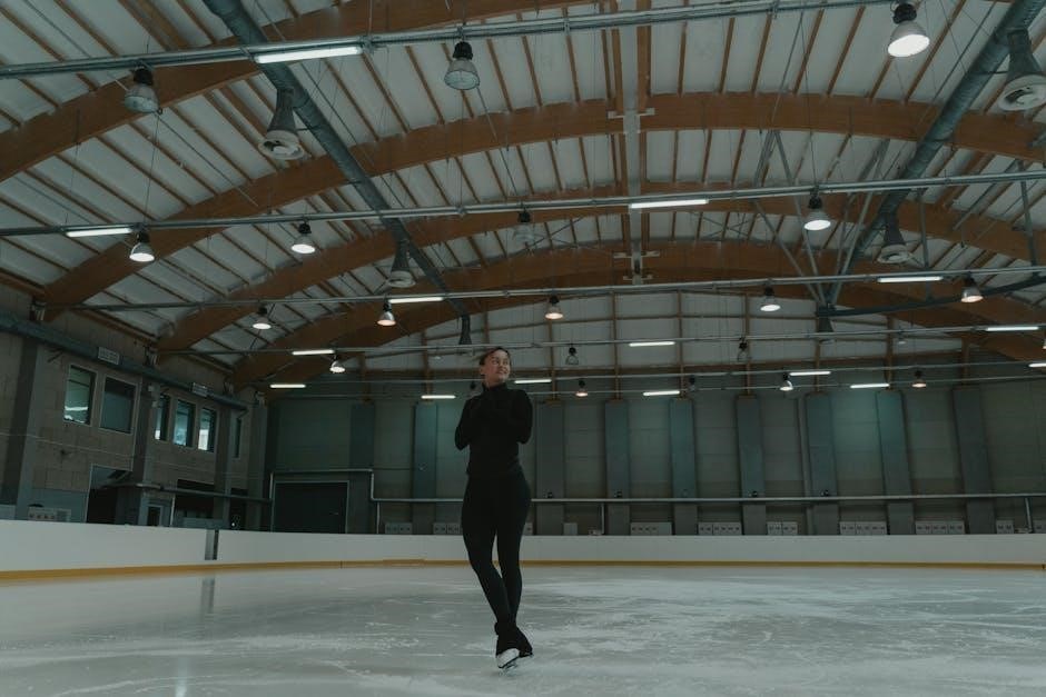
Balance and proprioception training is essential for restoring stability and preventing re-injury in peroneal tendonitis. Single-leg balance exercises are highly effective‚ where the individual stands on the affected leg‚ keeping the other foot lifted‚ and holds for 20-30 seconds. This improves ankle stability and reduces the risk of further strain. Wobble board exercises are another key component‚ involving standing on an unstable surface and performing controlled movements like tilting forward‚ backward‚ and side-to-side. These exercises enhance neuromuscular control and proprioception. Additionally‚ balance and reach exercises‚ where the individual stands on the injured leg and reaches for points around them‚ further challenge stability. These exercises should be performed gradually‚ starting with support if needed‚ and progressed as balance improves. Consistency in these practices helps restore functional stability and reduces the likelihood of recurrence. Regular practice‚ ideally 2-3 times daily‚ yields the best outcomes for recovery and long-term stability.
6.4 Range of Motion Exercises
Range of motion exercises are crucial for maintaining flexibility and reducing stiffness in the peroneal tendons and ankle joint. Ankle eversion is a key exercise‚ where the foot is turned outward to stretch the peroneal tendons. To perform this‚ sit with the affected leg crossed over the other‚ grip the foot‚ and gently turn the sole outward until a stretch is felt on the outside of the ankle. Hold for 15-20 seconds and repeat 3 times. Another effective exercise is the towel stretch‚ where a towel is looped around the toes‚ and gentle pulling stretches the calf and peroneal muscles. Additionally‚ ankle inversion and eversion exercises‚ performed while seated or standing‚ help restore natural movement. These exercises should be done 2-3 times daily to improve mobility and aid in recovery. Consistency is essential to prevent stiffness and promote healing.
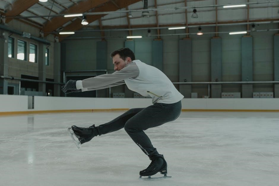
6.5 Dynamic Exercises
Dynamic exercises are essential for improving flexibility‚ strength‚ and functional movement in individuals with peroneal tendonitis. These exercises involve controlled‚ repetitive movements that mimic daily activities or sports-specific actions. A popular dynamic exercise is the high knees drill‚ where you march in place‚ lifting knees toward your chest to engage the peroneal tendons. Another effective exercise is calf hops‚ performed by hopping on one leg while keeping the knee straight‚ which strengthens the lower leg muscles. Lateral shuffles are also beneficial‚ as they target the peroneal tendons by moving side-to-side while keeping the feet low to the ground. These exercises should be done in 2-3 sets of 10-15 repetitions‚ gradually increasing speed and intensity as symptoms allow. Proper footwear and a smooth‚ even surface are recommended to ensure safety and effectiveness. Consistency in performing these exercises helps restore tendon function and reduces the risk of re-injury.
6.6 Calf and Ankle Mobilization
Calf and ankle mobilization exercises are crucial for restoring flexibility and reducing tension in the peroneal tendons. Start with a towel stretch: sit on the floor with your injured leg extended‚ loop a towel around your toes‚ and gently pull your foot back toward you. Hold for 15-30 seconds and repeat 3 times. For calf mobilization‚ perform a calf wall stretch by standing facing a wall with one leg extended behind you‚ keeping your heel on the ground. Lean forward until you feel a stretch in your calf‚ holding for 30 seconds and repeating 3 times on each side. These exercises improve ankle mobility and reduce stiffness‚ promoting recovery. Perform them 1-2 times daily‚ ensuring proper technique to avoid further injury.
6.7 Wobble Board Exercises
Wobble board exercises are excellent for improving balance‚ proprioception‚ and ankle stability‚ which are essential for recovering from peroneal tendonitis. Start by standing on the wobble board with your feet shoulder-width apart. Hold onto a chair for support if needed. Rock the board forward and backward 30 times‚ then side to side 30 times. Next‚ rotate the board in a circular motion‚ ensuring the edge remains in contact with the floor. Perform 3 sets of each movement. These exercises enhance dynamic stability and strengthen the muscles around the ankle‚ reducing the risk of further injury. Gradually increase the difficulty by closing your eyes or reducing support. Perform these exercises 2-3 times daily to improve balance and ankle function.
6.8 Single-Leg Balance Exercises
Single-leg balance exercises are crucial for improving stability and proprioception‚ which are essential for recovering from peroneal tendonitis. Stand on the injured leg‚ keeping the other foot lifted. Hold onto a chair for support if needed. Slowly raise the arch of the standing foot while keeping the toes on the floor. Hold this position for 10-15 seconds‚ then lower. Perform 3 sets of 10 repetitions. As balance improves‚ progress to standing without support. To increase difficulty‚ close your eyes or add gentle arm movements. This exercise strengthens the peroneal muscles and enhances ankle stability‚ reducing the risk of reinjury. Aim to practice this exercise 2-3 times daily for optimal results.
Preventing Peroneal Tendonitis
Preventing peroneal tendonitis involves proper footwear‚ gradual activity increases‚ and regular calf and peroneal muscle stretching. Strengthening exercises and maintaining optimal foot mechanics are also essential.
7.1 Proper Footwear
Wearing proper footwear is crucial in preventing peroneal tendonitis; Shoes should provide adequate support‚ especially for the arch and ankle‚ to reduce stress on the peroneal tendons. Avoid worn-out shoes or those with insufficient cushioning‚ as they can lead to poor foot mechanics. Opt for footwear with a sturdy heel counter and flexible sole‚ which helps stabilize the foot during movements. For athletes or individuals with high physical activity‚ motion control or stability shoes are recommended to minimize excessive pronation or supination. Additionally‚ replacing running shoes regularly‚ ideally every 300-500 miles‚ ensures continued support and cushioning. Proper footwear not only reduces the risk of developing peroneal tendonitis but also promotes overall foot health and functionality.
7.2 Calf and Peroneal Muscle Maintenance
Maintaining strong and flexible calf and peroneal muscles is essential for preventing peroneal tendonitis. Regular stretching and strengthening exercises help reduce tightness and imbalances that can strain the tendons. Focus on calf stretches‚ heel raises‚ and peroneal muscle exercises to improve flexibility and strength. Strengthening the muscles around the ankle joint ensures better support for the tendons‚ reducing the risk of overuse injuries. Additionally‚ incorporating exercises that target the peroneal muscles‚ such as ankle eversion and resistance band workouts‚ can enhance muscle endurance and stability. Proper maintenance also involves avoiding sudden increases in physical activity and ensuring adequate recovery time between workouts. By keeping these muscles strong and flexible‚ individuals can significantly lower their risk of developing peroneal tendonitis and promote long-term foot and ankle health.
7.3 Gradual Increase in Activity
A gradual increase in physical activity is crucial for preventing peroneal tendonitis and ensuring proper rehabilitation. Sudden spikes in exercise intensity or duration can overstrain the tendons‚ leading to inflammation and pain. By progressively loading the tendons‚ individuals allow them to adapt and strengthen without excessive stress. This approach minimizes the risk of re-injury and promotes long-term recovery. When resuming or increasing activity‚ it’s essential to monitor the body’s response and adjust accordingly. Incorporating rest periods and low-impact exercises initially can help the tendons acclimate to the demands. Avoiding aggressive training schedules and focusing on controlled‚ incremental progress ensures a safer and more effective path to full mobility and strength. Listening to the body and adhering to a structured plan are key to maintaining healthy tendons and preventing future issues.
Recovery and Rehabilitation Timeline
Recovery from peroneal tendonitis typically follows a phased approach‚ with the acute phase lasting 1-2 weeks‚ subacute phase 2-4 weeks‚ and return to activity phase 4-6 weeks.
8.1 Acute Phase
The acute phase of peroneal tendonitis recovery typically lasts 1-2 weeks‚ focusing on reducing pain and inflammation. During this period‚ rest‚ ice‚ compression‚ and elevation (RICE) are essential. Gentle exercises like heel raises‚ calf stretches‚ and ankle mobilization are introduced to maintain flexibility without aggravating the tendons. Pain management may include nonsteroidal anti-inflammatory medications. Patients are advised to avoid weight-bearing activities and use supportive footwear or braces to reduce stress on the tendons. Early mobilization is crucial to prevent stiffness‚ but exercises must be performed cautiously to avoid overloading the injured tissues. The goal is to minimize discomfort and prepare the tendons for more intensive rehabilitation in the subsequent phases.
8.2 Subacute Phase
The subacute phase of peroneal tendonitis rehabilitation typically lasts 2-4 weeks‚ focusing on progressive strengthening and mobility. During this stage‚ exercises such as single-leg balance‚ wobble board training‚ and resistance band eversion are introduced to enhance tendon strength and ankle stability. Gentle calf and peroneal stretching is continued to improve flexibility. Patients are encouraged to gradually resume weight-bearing activities‚ ensuring proper form to avoid re-injury. Dynamic exercises‚ such as heel-to-toe walking and lateral step-ups‚ are incorporated to restore functional movement. Pain management remains a priority‚ with a focus on reducing reliance on anti-inflammatory medications. The goal of this phase is to restore strength‚ balance‚ and mobility while preparing the tendons for more vigorous activities in the next phase of recovery.
8.3 Return to Activity Phase
The return to activity phase marks the final stage of rehabilitation‚ where patients gradually resume their normal activities and sports. This phase emphasizes functional exercises to restore full strength‚ flexibility‚ and coordination. Dynamic exercises‚ such as agility drills and sport-specific movements‚ are introduced to simulate real-life conditions. Patients are encouraged to perform activities like figure-eight runs and cone drills to enhance agility and balance. Pain should be minimal‚ and activities are progressed based on tolerance. It is crucial to maintain regular strengthening and stretching routines to prevent recurrence. Consultation with a healthcare provider is recommended before fully returning to high-impact activities to ensure complete recovery and minimize the risk of re-injury.